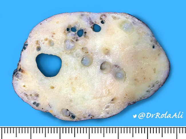
BR 4 - Blue dome cyst surrounded by normal breast tissue Benign cysts filled by serous fluid often have this blue color when viewed from the outside. Proximal tubule del3p clear cell renal cell carcinoma.

NaK ratio of 3 or less.
Fibrocystic change blue dome. The cut surface of fibrocystic change is characterized by variably-sized cysts scattered in fibrotic breast tissue. The cysts are filled with straw-colored to dark brown fluid. Given their appearance when intact the larger cysts are referred to as blue dome cysts.
A gross specimen shows two blue domed cysts separated by dense connective tissue. The term blue dome is applied to these cysts that are filled with dark fluid. The term fibrocystic change refers to benign breast changes caused by hormonally mediated exaggerated breast tissue response.
Common in premenopausal women. Disease subsides in post menopausal women as there is decrease in estrogen levels. Common age group is 30 -50 years.
Some of the larger cysts in fibrocystic change may have a bluish appearance from outside blue-domed cysts. The cyst lining is flattened or absent in some cases. In the center of this image cysts are lined by apocrine epithelium.
Note the focus of adenosis above it. Blue dome cysts. Based on gross appearance Type 1 cysts.
NaK ratio of 3 or less. Increased breast cancer risk. Associated with higher levels of estrogen melatonin epidermal growth factor and DHEA-S and lower levels of TGF-B2 than type 2 cysts Breast Cancer Res Treat 2007103331 Type 2 cysts.
Fibrocystic change usually bilateral breast tenderness seen in younger women that waxes and wanes with menstrual cycle blue dome cysts sclerosing adenosis apocrine metoplasia. However theres a Q on one of the newer obgyn forms where a woman has a unilateral painless mass that drains a serous brown fluid on FNA and the answer. The cysts range from 1 cm to 5 cm in diameter.
They are typically blue hence the nickname blue dome cysts. Epithelial hyperplasia is a proliferative fibrocystic change characterised by hyperplasia of the two epithelial layers of the terminal duct lobular units Pathology. Based on THAT apparently we were supposed to select fibroadenoma over fibrocystic change.
So I guess my question is essentially two-fold. Do we really have to know and distinguish among the histology of fibroadenoma and the multiple types of fibrocystic change including sclerosis adenosis cystic blue dome etc OR 2. You need to know fibrocystic change will show up with an unusual presentation on one of the obgyn forms.
Normally wed expect the standard bilateral breast tenderness in a young woman that waxes and wanes with the menstrual cycle. Wed see descriptors such as blue dome cysts or sclerosing adenosis or apocrine metaplasia. Grayish-white firm ovoid mass measuring 5 x 48 x 38 cm Fibrocystic Change Blue dome cyst Apocrine Metaplasia 46F CC.
Firm tender mass upper outer quadrant of left breast of 2 years duration No associated signs and symptoms but there was increase in size Excision biopsy was done Sclerosing Adenosis. ANSWER A BLUE DOMED CYST BLUE DOME CYSTS. Nonproliferative changes include fibrosis of the stroma and cystic dilation of the terminal ducts which when large may form blue-domed cysts.
BR 4 - Blue dome cyst surrounded by normal breast tissue Benign cysts filled by serous fluid often have this blue color when viewed from the outside. BR 5 - Extensive fibrocystic changes in serially sectioned formalin fixed breast tissue Cysts of various size are interspersed by dense fibrous tissue. This patient who was in a high risk category for breast carcinoma elected to.
Fibrocystic breast change is a common noncancerous condition that affects mostly premenopausal women. Fibrocystic breast changes encompass a wide variety of symptoms including breast tenderness or discomfort the sudden appearance or disappearance of palpable benign masses in the breast or lumpy free-moving masses in the breast. A 28-year-old female recently discovered a lump in her right breast.
She has no family history of breast cancer and her menstrual cycle began at 13 years of age. On exam you find a 1-cm movable mass in the right outerupper quadrant. There is no skin dimpling.
In fibrocystic change small cysts are surrounded by fibrous stroma. Large cysts can contain brown black fluid. Numerous variably sized cysts surrounded by foci of adenosis.
Some of the larger cysts in fibrocystic disease may have a bluish appearance from outside blue-domed cysts. The cyst lining is flattened or. The biopsy specimen at the lower right reveals a transected empty cyst.
Those on the left have unopened blue dome cysts. Microscopic detail of fibrocystic change of the breast revealing dilation of ducts producing microcysts and at right the wall of a large cyst with visible lining epithelial cells. A normal duct or acinus has a.
Fibrocystic breast changes can include simple cysts which are dilated and fluid-filled ducts. Papillary apocrine change or metaplasia. Now cysts in fibrocystic breast changes can be clear or blue-domed due to a light yellow fluid that gives the cyst a blue color when seen through the surrounding tissue.
Fibrocystic change occurs in multiple areas of both breasts. A dominant cyst or aggregate of fi brous connective tissue containing smaller cysts may manifest as a discrete mass prompting biopsy to exclude the possibility of cancer. The large cysts often contain dark fl uid that imparts a blue colorthe so-called blue-domed cysts of Bloodgood.
Blue domed cysts on gross breast tissue Apocrine metaplasia. Ductal hyperplasia and sclerosing adenosis calcified. Fibrocystic change of the breast includes a wide variety of changes of the breast ducts and stroma resulting in lumpy change of the breasts on physical examination.
Gross examination of breast tissue involved by fibrocystic change reveals dense white stromal tissue admixed with variably sized cysts that may have a brown or blue blue-dome cyst. Blue dome cyst Ductal ectasia Fibrocystic change Cracked Nipple Fistula Nipple Breast Disorder 35 Breast Disorder Fibrocystic change. Diffuse cystic mastopathy Fibrosclerosis Chronic cystic mastitis Mammary dysplasia Fibroadenosis microcyst of fibrocystic disease Breast cyst solitary cyst gross cyst Ductal ectasia.
Blue-dome cyst in Fibrocystic Change. What pathology is seen here. Fibrocystic change-Cysts are visible-fibrosis surrounds the cysts-sometimes the secretions win cysts will calcify.
What pathology is this. Apocrine Metaplasia in Fibrocystic Change pinkeosinophilic staining cells. Terms in this set 102 breast fibrocystic change blue dome cyst.
CMettrisomies distal tubule papillary renal cell carcinoma. Proximal tubule del3p clear cell renal cell carcinoma. Giant cell bacterial myocarditis.