The tail isnt just something which wags to show you theyre happy it serves a much bigger function. A Mid-sagittal T2W sequence of the cervical spinal cord revealing an intramedullary spinal lesion suggestive of gliosis or vasogenic oedema.

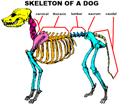
The spinal cord of dogs is divided into regions that correspond to the vertebral.
Husky spinal anatomy. Anatomy of the canine lumbar vertebrae and lumbosacral junction CT This anatomical module of the atlas of veterinary radiological anatomy vet-Anatomy covers the lumbar vertebrae lumbosacral joint sacrum and caudal vertebrae of the dog on a Computed Tomography CT and on 3D images of the lumbar spine and pelvic girdle. Spinal Column It consists of all the vertebrae and forms a part of the nervous system. Trachea The trachea is actually a tube that transports inhaled air to the lungs.
Esophagus It is the tube that connects the throat to the stomach thus aiding in transporting food for digestion. Larynx It houses the dogs vocal cords. Dog Tail Anatomy The tail of a dog serves many functions such as non verbal communication and as a rudder in water.
The tail isnt just something which wags to show you theyre happy it serves a much bigger function. They can be long short curly or flat. The tail is an extension of the spine so any injuries to the tail can be quite serious.
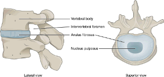
Intervertebral disk disease IVDD is a term for degeneration and protrusion of the intervertebral disk in dogs which leads to compression of. The Siberian husky is a medium-sized dog slightly longer than tall. Height ranges from 20 to 23 12 inches and weight from 35 to 60 pounds.
The Siberian husky has erect ears and eyes of brown to blue or maybe even one of each color. The neck is carried straight and the topline is level. The well-furred tail is carried up in a sickle or.
Official Standard of the Siberian Husky General Appearance. The Siberian Husky is a medium-sized working dog quick and light on his feet and free and graceful in. A Mid-sagittal T2W sequence of the cervical spinal cord revealing an intramedullary spinal lesion suggestive of gliosis or vasogenic oedema.
B Transverse computed tomography of the atlantoaxial junction revealing asymmetry of the articular facets of the axis. The maxilla is the bone that forms your upper jaw. The right and left halves of the maxilla are irregularly shaped bones that fuse together in the middle of the skull below the nose in an area known as the intermaxillary suture.
The maxilla is a major bone of the face. The Siberian Husky breed has an average lifespan of 12 to 14 years and are an ideal pet choice for lots of different people including families. However purebred Huskies do have a number of canine health problems that prospective owners should consider.
As with all animals its important to be aware of the common health concerns that plague Siberian Huskies since. The larynx is attached to the first ring of the trachea and is suspended ventral to the esophagus by the hyoid apparatus Figures 101-1 and 101-2Structurally it is formed by epiglottic thyroid cricoid sesamoid interarytenoid and paired arytenoid cartilages. 28 The epiglottis is the rostral most cartilage.
It is spade shaped with its. The workshop will focus specifically on sex after spinal cord injuries but the fundamental topics of communication and intimacy apply to everyone. The central nervous system includes the spinal cord and the brain.
The brain is divided into 3 main sectionsthe brain stem which controls many basic life functions the cerebrum which is the center of conscious decision-making and the cerebellum which is involved in movement and motor control. The spinal cord of dogs is divided into regions that correspond to the vertebral. A cats nervous system is a unique part of the feline anatomy.
The nervous system fully develops as the kitten ages barring any trauma or infection that can hinder this process. The central nervous system CNS is responsible for the brain and spinal cord messages the peripheral nervous system PNS affects muscles. A stout and short muscle lying posterior to the acromiodeltoid.
It lies along the lower border of the scapula and it passes through the upper arm across the upper end of muscles of the upper arm. It originates at the spine of the scapula and inserts at the deltoid ridge. Its action is to raise and rotate the humerus outward.
The muscular anatomy of a dog while serving the same purpose in a dog differs in structure and function from the muscular system in a human body. Just as the human muscular system is composed of units of tissue connected to the skeletal system skin and other muscles a dogs muscle anatomy is arranged in a similar fashion. The Back Pain Project.
Opening at 700 AM tomorrow. Call 203 424-2927 Get directions WhatsApp 203 424-2927 Message 203 424-2927 Contact Us Get Quote Find Table Place Order View Menu. Pain from upper abdominal structures passes via nerve fibers that run through the celiac plexus along the anterior surface of the aorta around the aorta posteriorly and then to splanchnic nerves on their way to the sympathetic chain and central nervous system.
Spondylosis also known as Spondylosis deformans is a degenerative disease that causes degenerative disks to form bone spurs between the vertebrae of the spine. This inhibits the flexibility and range of motion for the dog and it can also be extremely painful. Some people like to think of Spondylosis as arthritis of the spine.
The PNS has three basic functions. 1 conveying motor commands to all voluntary striated muscles in the body. 2 carrying sensory information about the external world and the body to the brain and spinal cord except visual information.
The optic nerves which convey information from the retina to the brain are in. Introduces students to functional human neuroanatomy of cerebral cortex basal ganglia thalamushypothalamus brains tern reticular formation cerebellum spinal cord motor system somatosensory system limbic system visual system auditoryvestibular system the blood supply of the nervous system cranial nerves and peripheral nerves autonomic nervous system. BackboneSpine Spinal ColumnVertebral Column.
Tail Caudal Appendage. Part Of NeckWindpipe LarynxPharynx. Upper Jaw Maxilla.
Lower Jaw Mandible. A Hoarse Voice Husky Huskily TerribleAwful Appalling. 441 Cervical spine OC1C7 35 442 Thoracic spine T1T13 35 443 Lumbar spine L1L7 35 444 Lumbosacral and sacroiliac joint 36 45 Equine vertebral column 37 451 Cervical spine OC1C7 37 452 Cervicothoracic junction C7T1 37 453 Thoracic spine T1T18 37 45 4 Lumbar spine L1L6 38.