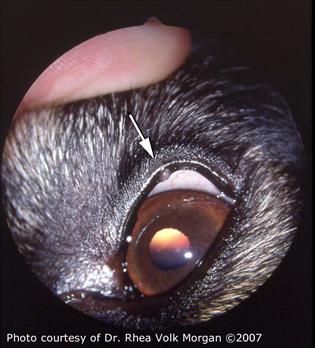
Meibomian gland adenoma and adenocarcinomaThese masses arise from Meibomian glands which are specialized glands that line the upper and lower eyelids. Meibomian gland adenoma and adenocarcinomaThese masses arise from Meibomian glands which are specialized glands that line the upper and lower eyelids.

-They are rubbing on the eye and bothering the dog.
Meibomian gland cyst adenoma. Meibomian Gland Adenomas MGA are benign age related eyelid tumors which result from the accumulation of glandular material. If they become large enough MGAs can cause irritation to the cornea and conjunctiva and may reduce the normal ability to blink. Over a period of 1 year a 74-year-old man slowly developed a painless left upper eyelid intratarsal mass.
The skin was movable over the lesion. At surgery a well-circumscribed yellow-white partially cystic tumor was encountered. Histopathologically it was composed of a.
Meibomian gland tumors are tiny slow-growing tumors that form in the meibomian glands of the eyelids. Bumps in or near the eyes can be uncomfortable because they scratch the cornea or prevent the eye from shutting properly. If you notice a growth on your dogs eyelid it could be whats known as a meibomian gland cyst or chalazion.
Both dog and cat eyes may be affected by these but they are more common in dogs. This is a tiny little growth that originates from the eyelid. In almost every case the growth is not cancerous.
-They are getting larger than 14 of the eyelid. -They are rubbing on the eye and bothering the dog. -They are ulcerating and bleeding.
Unfortunately the only way to get rid of a meibomian gland adenoma is to have it surgically removed. Meibomian cyst develops when there is a problem with the eyelid glands. As meibomian glands secrete the lubricating sebum problem arises as the glands are blocked because of some underlying condition such as narrowed opening of the glands or from accumulation of hardened secretions.
A meibomian cyst commonly referred to as chalazion is a non-infectious inflammatory disease of the sebaceous glands on the eyelids that presents as an enlarging lump over the course of days or weeks without resolution to topical antimicrobial therapy. Acne rosacea and seborrheic dermatitis increase the risk for meibomian cysts. Blurred vision may be encountered with.
The edges of the eyelids for example have tiny glands called meibomian glands containing cells that produce secretions to lubricate the eye. If these cells overmultiply they develop into benign tumors called meibomian gland adenomas non-cancerous or meibomian gland adenocarcinomas a less common malignant tumor. Meibomian gland adenoma and adenocarcinomaThese masses arise from Meibomian glands which are specialized glands that line the upper and lower eyelids.
Meibomian glands secrete oily substances that help keep the tear film healthy. Masses arising from these glands are often seen protruding from the eyelid margin. Sebaceous ductal adenoma Meibomian ductal adenoma.
Haphazard arrangement of predominantly ducts admixed with fewer basaloid reserve cells and sebocytes Sebaceous carcinoma Meibomian carcinoma. Variable cytoplasmic lipid content. Meibomian Adenoma Meibomian Ductal Adenoma Meibomian Epithelioma Meibomian Carcinoma Description Meibomian glands are a special kind of sebaceous glands located on the periphery of the eyelid.
They are responsible for the supply of sebum which prevents the tear film from evaporating. The most common types of tumors appear as neoplasia of the Meibomian gland the primary oil producing glands located in the eyelid margin. There are dozens of these glands in each eyelid and the origin of these tumors is usually either the duct linings epithelioma or the ascini adenoma that grow as multilobulated pink to grey well vascularized Figure 1.
Oil glands withing the eyelid whose duct opens onto the eyelid. Secretions supply the outer portion of the tear film preventing rapid tear evaporation. These glands can be visualized using IR technique to determine if there is meibomian gland dysfunction MGD.
These tumors are tiny slow-growing tumors that form in the meibomian glands of the eyelids. Meibomian glands are sebaceous glands that provide an oily secretion to stabilize the tear film over the cornea Common in older dogs meibomian gland tumors are usually benign but a small percentage of them are carcinomas that can metastasize into lymph nodes. Chalazion is a lipogranuloma of one of the meibomian glands of the tarsal plate.
They arise relatively rapidly over a period of a few days often with inflammation and discomfort. They can progress to form a chronic pea-sized firm nodule in the lid Figure 13The initial treatment is to hot compress the involved lid frequently in the first few days in hopes of opening the. Using a wedge resection we removed a large Meibomian cyst from a dogs upper lid.
It goes without saying that these videos are intended for veterinary prof. Thirteen-year-old spayed female Labrador retriever with a meibomian gland adenoma. Note the mass arising from the superior eyelid erupting through the eyelid margin to the palpebral conjunctiva.
The mass is causing local irritation characterized by conjunctival hyperemia and epiphora. Surgical correction which was curative with a 4-sided. A novel meibomian gland dysfunction MGD model induced by the injection of complete Freunds adjuvant CFA in rabbits was developed to facilitate the understanding of the pathophysiology of MGD with meibomitis.
In addition we sought to evaluate. Most common ocular neoplasm in dogs histological appearance consistent with sebaceous adenomas. Meibomian gland epitheliomas are composed of densely packed sheets of basal reserve cells forming well defined lobules.
They are sebaceous glands which are also found in other areas of the skin. Tumors in the Meibomian glands result in bumps along the inside or edges of the eyelids. They may pop outward or turn inward and rub on the cornea.
Meibomian gland tumors in dogs are usually benign non-cancerous so they dont typically spread or move to other areas of the body. What are meibomian gland tumours. These are tumours of the meibomian glands of the eyelids.
These are common in older dogs and start as small bumps at the margin of the upper and lower eyelids. Many of these stay small 2 - 3mm and do not continue to grow further so there is never any rush to have them removed. Meibomian gland cysts in dogs are tiny little nodules that can form in whats known as the third eye.
The third eye aka nictitating membrane is the tissue you see in the corner of the eye. If you watch your dog blink youll see movement of that membrane. Will My Dog Go Blind.
Meibomian cysts or tumors occur on or under the eyelid margin.